


2012/03/13
Mad rabbit disease
The ability of prions to infect some species and not others is determined by the transmission barrier. This unexplained phenomenon led to the belief that certain species were not susceptible to transmissible spongiform encephalopathies (TSEs) and therefore represented negligible risk to human health if consumed. Using the Protein Misfolding Cyclic Amplification (PMCA) technique, we were able to overcome the species barrier in rabbits, which have been classified as TSE resistant for four decades. Rabbit brain homogenate, either unseeded or seeded in vitro with disease related prions obtained from different species, was subjected to serial rounds of PMCA. De novo rabbit prions produced in vitro from unseeded material were tested for infectivity in rabbits, with one of three intra-cerebrally challenged animals succumbing to disease at 766 days and displaying all the characteristics of a TSE, thereby demonstrating that leporids are not resistant to prion infection. Material from the brain of the clinically affected rabbit containing abnormal prion protein resulted in a 100% attack rate after its inoculation in transgenic mice over-expressing rabbit PrP. Transmissibility to rabbits (> 470 days) has been confirmed at least in 2 out of 10 rabbits after intra-cerebral challenge. Despite rabbits no longer being able to be classified as resistant to TSEs an outbreak of 'mad rabbit disease' is unlikely.
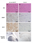
Figure: Histological and immunohistochemical studies. Sections of midbrain from a rabbit challenged with de novo RaPrPSc (A, C, E and G) and a negative control unchallenged rabbit (B, D, F and H). Hematoxylin and eosin (HE) stained sections showed vacuolation, predominantly in the neuropil of the brain of the challenged rabbit (A) but none in the negative control animal (B). Immunohistochemistry using antibodies to GFAP (glial fibrilary acidic protein) and abnormal prion protein (2G11) showed respectively astrocytosis (C, brown pigment) and positive labeling of PrPSc (E, brown pigment) in the brain of the challenged rabbit. Neither astrocytosis (D) nor PrPSc (F) was found in the brain of the negative control rabbit. PET-Blot analysis using SAF84 for PrPSc was positive in the challenged rabbit (G, black pigment) but absent in the negative control (H).
LINK: PNAS
LINK: El País
LINK: El Correo
LINK: Youtube
See a large version of the first picture